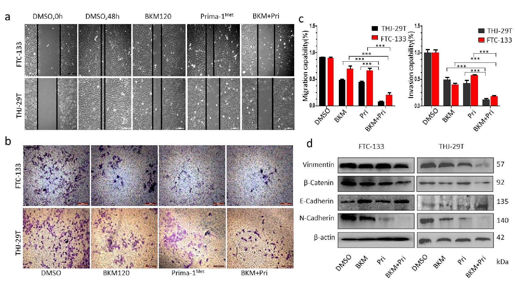
Fig. 2. Effect of the combination treatment of NVP-BKM120 and Prima-1Met on migration and invasion of thyroid cancer cells. (a) Cell migration was analyzed by wound healing assay. THJ-29T and FTC-133 cells were grown to 70-80% confluency. The cell monolayers were wounded with a sterile pipette tip, and washed with medium to remove detached cells from the plates. Then the cells were left either untreated or treated with BKM120 (1 µM), Prima-1Met (30 µM), or the two in combination. After 48 hours, the wound gap was observed and photographed. *P<0.05, significant difference between the BKM120 + Prima-1Met treated group and the BKM120 or Prima-1Met treated groups. (b) FTC-133 and THJ-29T cancer cells were subjected to Matrigel invasion assay and photographed (magnification 20×, scale bar 100 µm). (c) Left: Migration capability of cells in (a) was calculated; Right: invasion capability of cells in (b) was calculation. The data were presented as mean ± SD of three independent experiments. ***P<0.005, significant difference between the treatment and control groups. (d) Protein levels of Vimentin, ß-catein, E-Cadherin and N-Cadherin were detected after treatment with BKM120 (1 µM), Prima-1Met (30 µM) or the two in combination. *P<0.05, significant difference between treatment group and DMSO control group. **P<0.05, significant difference between combination treatment group and single-agent treatment group. All the experiments were repeated 3 times, and the data were shown as mean ± SD.